6.5.1 State that the nervous system consists of the central nervous system (CNS) and peripheral nerves, and is composed of cells called neurons that carry rapid electrical impulses
Neurons are cells that are specialised for the conduction of nerve impulses and serve as the fundamental unit of the nervous system
The nervous system can be divided into two main parts:
- Central Nervous System (CNS): Made up of the brain and the spinal cord
- Peripheral Nervous System (PNS): Made of peripheral nerves which link the CNS with the body's receptors and effectors
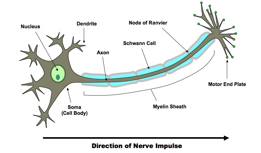
6.5.2 Draw and label a diagram of the structure of the motor neuron
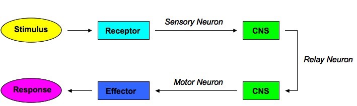
6.5.3 State that nerve impulses are conducted from receptors to the CNS by sensory neurons, within the CNS by relay neurons, and from the CNS to effectors by motor neurons
There are three main types of neurons in the nervous system:
- Sensory Neurons: Conduct nerve impulses from receptors to the CNS (afferent pathway)
- Relay Neurons: Conduct nerve impulses within the CNS (also called interneurons or connector neurons)
- Motor Neurons: Conduct nerve impulses from the CNS to effectors (efferent pathway)
The Stimulus-Response Pathway
6.5.4 Define resting potential and action potential (depolarisation and repolarisation)
Resting Potential: The charge difference across the membrane when a neuron is not firing (-70 mV), as maintained by the sodium-potassium pump
Action Potential: The charge difference across the membrane when a neuron is firing (about 30 mV)
Depolarisation: The change from a negative resting potential to a positive action potential (caused by opening of sodium channels)
Repolarisation: The change from a positive action potential back to a negative resting potential (caused by opening of potassium channels)
6.5.5 Explain how a nerve impulse passes along a non-myelinated neuron
Generation of a Resting Potential
- The sodium-potassium pump (Na+/K+ pump) maintains the electrochemical gradient of the resting potential (-70 mV)
- It is a transmembrane protein that uses active transport to exchange Na+ and K+ ions across the membrane (antiport mechanism)
- It expels 3 Na+ ions for every 2 K+ ions admitted (in addition, some of the K+ ions will leak back out of the cell)
- This makes the inside of the membrane relatively negative when compared to the outside (-70 mV = resting potential)
Transmission of an Action Potential
- Sodium and potassium channels in nerve cells are voltage-gated, meaning they can open and close depending on the voltage across the membrane
- In response to a signal at a sensory receptor or dendrite, sodium channels open and sodium enters the neuron passively
- The influx of sodium (Na+ in) causes the membrane potential to become positive (depolarisation)
- If a sufficient change in membrane potential is achieved (threshold potential), adjacent voltage-gated sodium channels open, generating a wave of depolarisation (action potential) that spreads down the axon
- The change in membrane potential also activates voltage-gated potassium channels, causing potassium to exit the neuron passively
- The efflux of potassium (K+ out) causes the membrane potential to become negative again (repolarisation)
- Before the neuron can fire again, the original distribution of ions (Na+ out, K+ in) must be re-established by the Na+/K+ pump
- The inability to propagate another action potential during this time (refractory period) ensures nerve impulses only travel in one direction
![]() Saltatory Conduction
Saltatory Conduction
Generation of an Action Potential

6.5.6 Explain the principles of synaptic transfer
- The junction between two neurons is called a synapse, it forms a physical gap between the pre-synaptic and post-synaptic neurons
- An action potential (electrical signal) cannot cross the synaptic gap, so it triggers the release of chemicals (neurotransmitters) to continue the signal
Chemical Transfer Across Synapses
- When an action potential reaches the axon terminal, it triggers the opening of voltage-gated calcium channels
- Calcium ions (Ca2+) diffuse into the cell and promote the fusion of vesicles (containing neurotransmitters) with the plasma membrane
- The neurotransmitters are released from the axon terminal by exocytosis and cross the synaptic cleft
- Neurotransmitters bind to appropriate neuroreceptors on the post-synaptic membrane, opening ligand-gated channels
- Excitatory neurotransmitters (e.g. noradrenaline) open ligand-gated sodium channels (depolarisation)
- Inhibitory neurotransmitters (e.g. GABA) open ligand-gated potassium or chlorine channels (hyperpolarisation)
- The combination of chemical messengers received by dendrites determines whether the threshold is reached for an action potential in the post-synaptic neuron
- Neurotransmitter molecules released into the synapse are either recycled (by reuptake pumps) or degraded (by enzymatic activity)
Overview of Synaptic Transfer
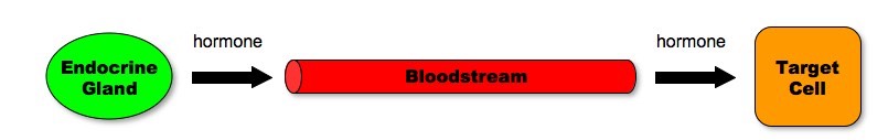
6.5.7 State that the endocrine system consists of glands that release hormones that are transported in the blood
An endocrine gland is a ductless gland in the body that manufactures chemical messengers called hormones and secretes them directly into the blood
Hormones act on distant sites (target cells) and tend to control slow, long-term activities such as growth and sexual development
![]() Endocrine System
Endocrine System
6.5.8 State that homeostasis involves maintaining the internal environment between limits, including blood pH, carbon dioxide concentration, blood glucose concentration, body temperature and water balance
Homeostasis is the tendency of an organism or cell to maintain a constant internal environment within tolerance limits
Internal equilibrium is maintained by adjusting physiological processes, including:
- Body temperature (normally 36 - 38°C)
- Blood pH (normally 7.35 - 7.45)
- Carbon dioxide concentration (normally 35 - 45 mmHg)
- Blood glucose concentration (normally 75 - 95 mg / dL)
- Water balance (varies with individual body size)
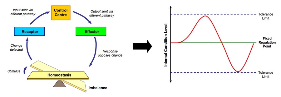
6.5.9 Explain that homeostasis involves monitoring levels of variables and correcting changes in levels by negative feedback mechanisms
- Most homeostatic control mechanisms operate through a negative feedback loop
- When specialised receptors detect a change in an internal condition, the response generated will be the opposite of the change that occurred
- When levels have returned to equilibrium, the effector ceases to generate a response
- If levels go too far in the opposite direction, antagonistic pathways will be activated to restore the internal balance
Negative Feedback Loop
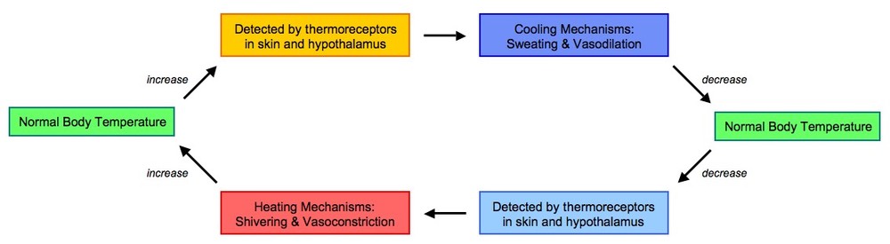
6.5.10 Explain the control of body temperature, including the transfer of heat in blood, and the roles of the hypothalamus, sweat glands, skin arterioles and shivering
Animals capable of temperature regulation within a given range are called homeotherms and maintain a constant body temperature through a negative feedback loop
- The hypothalalmus acts as a control centre in thermoregulation by detecting fluctuations in body temperature
- The skin also possesses thermoreceptors and relays this information to the hypothalamus, which coordinates corrective measures
When body temperature rises, the following cooling mechanisms may occur:
- Vasodilation: The skin arterioles dilate, bringing blood into closer proximity to the body surface and allowing for heat transfer (convective cooling)
- Sweating: Sweat glands release sweat, which which is evaporated at the cost of latent heat in the air, thus cooling the body (evaporative cooling)
When body temperature falls, the following heating mechanisms may occur:
- Vasoconstriction: The skin arterioles constrict, moving blood away from the surface of the body, thus retaining the heat carried within the blood
- Shivering: Muscles begin to shake in small movements, expending energy through cell respiration (which produces heat as a by-product)
Other mechanisms through which homeotherms may regulate their body temperature include:
- Piloerection: Animals with furry coats can make their hair stand on end (piloerection), trapping pockets of warm air close to the body surface
- Behavioural responses: Animals may physically respond to environmental conditions in a bid to regulate temperature (e.g. bathing, burrowing, etc.)
Thermoregulation by the Nervous System
6.5.11 Explain the control of blood glucose concentration, including the roles of glucagon, insulin and the alpha and beta cells in the pancreatic islets
The body requires volumes of glucose in order to make ATP, however the amount of ATP demand will fluctuate according to need and thus the body regulates its release of glucose into the bloodstream as high levels of glucose in the bloodstream can damage cells (creates hypertonicity)
- Two hormones, insulin and glucagon, are responsible for controlling blood glucose concentration (they have antagonistic functions)
- These hormones are released from different groups of cells with pancreatic pits (called the islets of Langerhans) and act principally on the liver
When blood glucose levels are high (e.g. after feeding):
- Insulin is released from beta cells in the pancreas and causes a decrease in blood glucose concentration
- This may involve stimulating glycogen synthesis in the liver (glycogenesis), promoting glucose uptake into the liver and adipose tissue or increasing the rate of glucose breakdown (increase cell respiration)
When blood glucose levels are low (e.g. after strenuous exercise):
- Glucagon is released from alpha cells in the pancreas and cause an increase in blood glucose concentration
- This may involve stimulating glycogen breakdown in the liver (glycogenolysis), promoting glucose release from the liver and adipose tissue or decreasing the rate of glucose breakdown (decrease cell respiration)
Blood Glucose Regulation by the Endocrine System

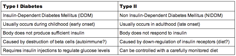
6.5.12 Distinguish between type I and type II diabetes