6.2.1 Draw and label a diagram of the heart showing the four chambers, associated blood vessels, valves and the route of the blood through the heart
Valves and Direction of Blood Flow Heart Chambers and Vessels

6.2.2 State that coronary arteries supply heart muscle with oxygen and nutrients
- The heart is a muscle that must continually contract in order to pump blood around the body
- Coronary arteries form a network of vessels around the heart and supply the cardiac tissue with oxygen and nutrients (i.e. glucose)
- These are required to produce the necessary energy via aerobic respiration - if a coronary artery is blocked, a heart attack may occur
6.2.3 Explain the action of the heart in terms of collecting blood, pumping blood and opening and closing valves
Blood returning from all parts of the body (except lungs) enter the right atrium via the vena cava - this blood is relatively deoxygenated
The blood passes from the right atrium to the right ventricle and then via the pulmonary artery to the lungs (where blood is reoxygenated)
The blood returns to the left atrium via the pulmonary vein and passes through the left ventricle to the aorta, where it is pumped around the body
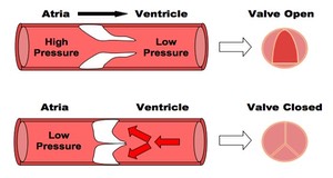
The heart valves maintain the one-way flow of blood:
- When the atria contract, atrioventricular (AV) valves open
- Blood flows from the atria and into the ventricles
- When the ventricles contract, the AV valves close and semilunar valves open
- This forces blood out of the ventricles and into the arteries
- As arterial pressure rises, the semilunar valves close, ensuring the one-way flow of blood
6.2.4 Outline the control of the heartbeat in terms of myogenic muscle contraction, the role of the pacemaker, nerves, the medulla of the brain and epinephrine (adrenaline)
- The contraction of the heart tissue (myocardium) is myogenic, meaning the signal for cardial contraction arises within the heart muscle itself
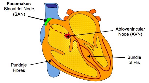
- Within the wall of the right atrium are a specialised plexus of nerves called the sinoatrial node (SAN)
- The sinoatrial node initiates contraction of the cardiace muscle and acts as a pacemaker, regulating normal sinus rhythm
- It stimulates atria to contract and, when excitation reaches the junction between atria and ventricles, stimulates another node (atrioventicular node)
- The atrioventricular node (AVN) sends signals via the Bundle of His to Purkinje fibres, which cause ventricular contraction
- This sequence always ensures their is a delay between atrial and ventricular contractions, resulting in two heart sounds ('lub dub')
Myogenic Control of the Heart Beat
The pacemaker is under autonomic control from the brain, specifically the medulla oblongata (brain stem)
- Sympathetic nerves speed up heart rate by releasing a neurotransmitter (noradrenaline) to increase the rate of myocardial contraction
- Parasympathetic nerves splow down heart rate by releasing a neurotransmitter (acetylcholine) to decrease the rate of myocardial contraction
- Additionally, the heart rate may be increased by the chemical release of the hormone adrenaline into the blood (from the adrenal gland)
6.2.5 Explain the relationship between the structure and function of arteries, capillaries and veins
Arteries
- Arteries carry blood at high pressure (80 - 120 mm Hg)
- They have a narrower lumen (to maintain high pressure) surround by a thick wall made of two layers
- The middle layer (tunica media) contains muscle and elastin to help maintain pulse flow (it can contract and stretch)
- The outer layer (tunica adventitia) contains collagen prevents the artery rupturing due to the high pressure blood flow
Veins
- Veins carry blood under low pressure (<10 mm Hg)
- They have a very wide lumen (keeps pressure low and allows greater flow of blood)
- The walls of tissue surrounding the vein are thin (blood is not travelling in rhythmic pulses)
- They have valves to prevent blood pooling at extremities (arteries do not have valves)
Capillaries
- Capillaries are involved with material and gas exchange with the surrounding body tissue
- Blood pressure in the capillaries is relatively low (~15 mm Hg) and they have a very small diameter (~5 micrometers wide)
- Their wall is made up a a single layer of cells to allow for ease of diffusion
- Capillaries may contain pores to aid the transport of material
Structure of Blood Vessels

6.2.6 State that blood is composed of plasma, erythrocytes, leukocytes (phagocytes and lymphocytes) and platelets
There are four main components to blood:
- Plasma - the fluid medium of the blood
- Erythrocytes - red blood cells (involved in oxygen transport)
- Leukocytes - white blood cells, such as phagocytes (non-specific immunity) and lymphocytes (specific immunity)
- Platelets - responsible for blood clotting (haemostasis)
6.2.7 State that the following are transported by blood: nutrients, oxygen, carbon dioxide, hormones, antibodies, urea and heat
The following things are transported by blood:
- Nutrients (e.g. glucose)
- Antibodies
- Carbon dioxide
- Hormones
- Oxygen
- Urea
- Heat (not a molecules, unlike all the others)