11.4.1 Annotate a light micrograph of testis tissue to show the location and function of interstitial cells (Leydig cells), germline epithelium cells, developing spermatozoa and Sertoli cells
Testis Tissue

- The testes are composed of seminiferous tubules which produce sperm
- Each tubule is surrounded by a basement membrane which is lined by germline epithelium cells
- The germline epithelium will divide by mitosis to make spermatogonia (which divide by meiosis to make spermatozoa)
- The developing spermatozoa are nourished by Sertoli cells
- Outside of the tubules are blood capillaries and interstitial cells (Leydig cells), which produce the male sex hormone, testosterone
11.4.2 Outline the processes involved in spermatogenesis within the testes, including mitosis, cell growth, the two divisions of meiosis and cell differentiation
- Spermatogenesis describes the production of spermatozoa (sperm) in the seminiferous tubules of the testes
- The first stage of sperm production requires the division of germline epithelium by mitosis
- These cells (spermatogonia) then undergo a period of growth
- This is followed by two meiotic divisions that result in four haploid daughter cells
- These haploid cells then differentiate to form sperm cells
- The developing sperm cells are nourished throughout by the Sertoli cells
Overview of Spermatogenesis

11.4.3 State the role of LH, testosterone and FSH in spermatogenesis
LH: Stimulates the interstitial cells (Leydig cells) to produce testosterone
FSH: Stimulates the (first) meiotic division of spermatogonia
Testosterone: Stimulates the (second) meiotic division of spermatogonia and the maturation of spermatozoa through differentiation
11.4.4 Annotate a diagram of the ovary to show the location and function of germline epithelium, primordial follicles, mature follicles and secondary oocyte
Structure of the Ovary

- The ovary contains follicles in various stages of development
- Egg cells within primordial follicles have been arrested in prophase I and have yet to undergo meiotic division
- Egg cells within mature follicles have begun meiotic division and are released from the ovary as secondary oocytes (arrested in prophase II)
- The ruptured follicle develops into a corpus luteum that will, in time, degenerate into a corpus albicans
- The germline epithelium functions as an epithelial layer separating ovarian tissue from the rest of the body - it is not involved in oocyte development
11.4.5 Outline the processes involved in oogenesis within the ovary, including mitosis, cell growth, the two divisions of meiosis, the unequal division of cytoplasm and the degeneration of polar body
- Oogenesis describes the production of female gametes (ova) within the ovary
- The process begins during foetal development, when a large number of cells (oogonia) are formed by mitosis before undergoing a period of growth
- These cells begin meiosis but are arrested in prophase I until puberty
- At puberty, some follicles continue to develop each month is response to FSH secretion
- These follicles complete the first meiotic division to form two cells of unequal size
- The cell with less cytoplasm is a polar body (which degenerates), while the larger cell forms a secondary oocyte
- The secondary oocyte begins the second meiotic division but is arrested in prophase II (until fertilisation)
- It is released from the ovary (ruptured follicle develops into corpus luteum) and, if fertilisation occurs, will complete meiosis
- The second meiotic division will produce an ovum and a second polar body
Overview of Oogenesis

11.4.6 Draw and label a diagram of a mature sperm and egg

11.4.7 Outline the role of the epididymis, seminal vesicle and prostate gland in the production of semen
Epididymis
- Testicular fluids are removed, concentrating the sperm
- Sperm mature and develop the ability to swim
Seminal Vesicle
- Adds nutrients (including fructose) for respiration
- Secretes prostaglandins, causing contractions to the female system and helping sperm move towards the egg
Prostate Gland
- Secretes alkaline fluid which neutralises vaginal acids (changes pH from 4 to 6 which aids sperm motility)
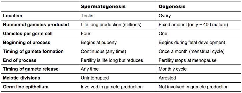
11.4.8 Compare the processes of spermatogenesis and oogenesis, including the number of gametes and the timing of formation and release of gametes
Similarities:
- Both processes result in the formation of haploid gametes
- Both processes involve mitosis, growth and meiosis
Differences:
11.4.9 Describe the process of fertilisation, including the acrosome reaction, penetration of the egg membrane by a sperm and the cortical reaction
- When the sperm enters the female reproductive tract, biochemical changes to the sperm occur in the final part of its maturation (capacitation)
- The sperm is attracted to the egg due to the release of chemical signals from the secondary oocyte (chemotaxis)
- Fertilisation generally occurs in the oviduct (fallopian tube)
- To enter the egg membrane, the sperm must penetrate the protective jelly coat (zona pellucida) surrounding the egg via the acrosome reaction
- The acrosome vesicle fuses with the jelly coat and releases digestive enzymes which soften the glycoprotein matrix
- The membrane of the egg and sperm then fuse and the sperm nucleus (and centriole) enters the egg
- To prevent other sperm from penetrating the fertilised egg (polyspermy), the jelly coat undergoes biochemical changes via the cortical reaction
- The cortical granules release enzymes that destroy the sperm-binding proteins on the jelly coat
- Now fertilised, the nucleus of the secondary oocyte completes meiosis II and then the egg and sperm nuclei fuse to form a diploid zygote
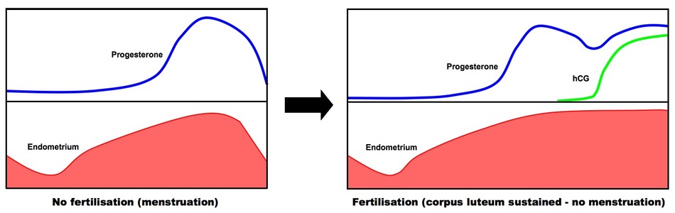
11.4.10 Outline the role of hCG in early pregnancy
- The endometrium is a blood-rich environment in which an implanted zygote can grow and it is sustained by the hormone progesterone
- If progesterone levels aren't maintained (i.e. the corpus luteum degenerates), then the endometrium will be sloughed away (menstruation)
- A fertilised zygote develops into a blastocyst that secretes human chorionic gonadotrophin (hCG)
- hCG maintains the corpus luteum post-ovulation so that the blastocyst can remain embedded in the endometrium and continue to develop
- Gradually the placenta develops and produces progesterone (at around 8 - 10 weeks), at which point the corpus luteum is no longer needed
Role of hCG in Early Pregnancy
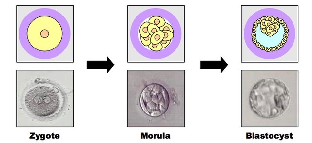
11.4.11 Outline early embryo development up to the implantation of the blastocyst
- After fertilisation, the zygote undergoes several mitotic divisions to create a solid ball of cells called a morula (at around 4 days)
- Unequal divisions beyond this stage cause a fluid-filled cavity to form in the middle - this makes a blastocyst (at around 5 days)
- The blastocyst consists of:
- An inner mass of cells (this will develop into the embryo)
- An outer layer called the trophoblast (this will develop into the placenta)
- A fluid filled cavity (called the blastocoele)
- These developments all occur as the developing embryo is moving from the oviduct to the uterus
- When the blastocyst reaches the uterus, it will embed in the endometrium (implantation)
Early Embryo Development
11.4.12 Explain how the structure and function of the placenta, including its hormonal role in secretion of estrogen and progesterone, maintain pregnancy
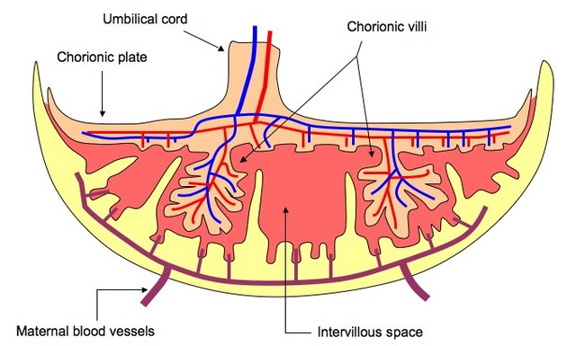
Structure and Function
- The placenta is a disc-shaped structure that nourishes the developing embryo
- It is formed from the development of the trophoblast upon implantation and eventually invades the uterine wall
- The umbilical cord connects the fetus to the placenta and maternal blood pools via open ended arterioles into intervillous spaces (lacunae)
- Chorionic villi extend into these spaces and facilitate the exchange of materials between the maternal blood and fetal capillaries
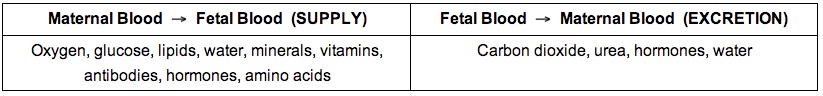
- Nutrients, oxygen and antibodies will be taken up by the fetus, while carbon dioxide and waste products will be removed
- The placenta is expelled from the uterus after childbirth
Hormonal Role
- The placenta also takes over the hormonal role of the ovary (at around 12 weeks)
- Estrogen stimulates growth of the muscles of the uterus (myometrium) and the development of the mammary glands
- Progesterone maintains the endometrium, as well as reduces uterine contractions and maternal immune response (no antibodies against fetus)
- Both estrogen and progesterone levels drop near time of birth
Structure of the Placenta
11.4.13 State that the fetus is supported and protected by the amniotic sac and amniotic fluid
- The fetus develops in a fluid-filled space called the amniotic sac
- Amniotic fluid is largely incompressible and good at absorbing pressure, and so protects the child from impacts to the uterine wall
- The fluid also creates buoyancy so that the fetus does not have to support its own body weight while the skeletal system develops
- Finally, amniotic fluid prevents dehydration of the tissues, while the amniotic sac provides an effective barrier against infection
11.4.14 State that materials are exchanged between the maternal and fetal blood in the placenta
The fetus relies on the exchange of materials across the placental wall to grow and develop:
11.4.15 Outline the process of birth and its hormonal control, including the changes in progesterone and oxytocin levels and positive feedback
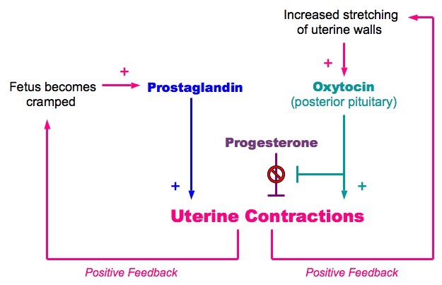
- The process of childbirth is called parturition and is controlled by the hormone oxytocin
- After nine months, the fetus is fully grown and takes up all available space in the uterus, stretching the walls of the uterus
- This causes a signal to be sent to the brain, releasing oxytocin from the posterior pituitary
- Oxytocin inhibits progesterone, which was inhibiting uterine contractions
- Oxytocin also directly stimulates the smooth muscle of the uterine wall to contract, initiating the birthing process
- The contraction of the uterine wall causes further stretching, which triggers more oxytocin to be released (causing even more contraction)
- Additionally, the fetus responds to the cramped conditions by releasing prostaglandins which cause further myometrial contractions
- As the stimulus causing oxytocin release is increased by the effects of oxytocin, this creates a positive feedback pathway
- Contractions will stop when labour is complete and the baby is birthed (no more stretching of the uterine wall)
The Hormonal Control of Child Birth