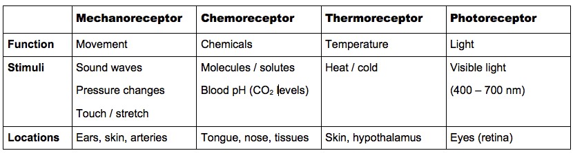
E.2.1 Outline the diversity of stimuli that can be detected by human sensory receptors
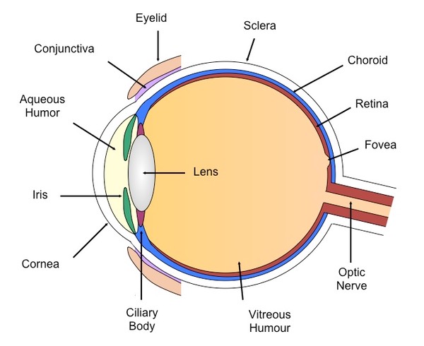
E.2.2 Label a diagram of the structure of the human eye
E.2.3 Annotate a diagram of the retina to show the cell types and the direction in which light moves

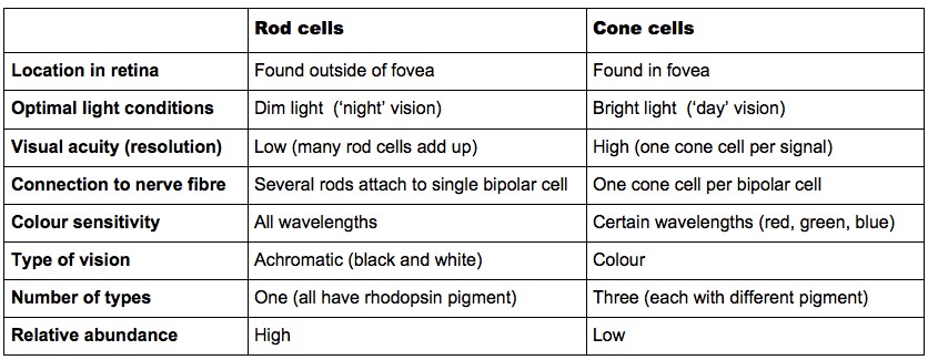
E.2.4 Compare rod and cone cells
Similarities:
- Both types of cell are photoreceptors
- Both types of cell are found in the retina
Differences:
E.2.5 Explain the processing of visual stimuli, including edge enhancement and contralateral processing
Photoreceptors (rod and cone cells) in the retina convert light into nerve impulses
The impulses pass to bipolar cells, which relay the signal to the optic nerve (via ganglion cells)
Edge Enhancement
Signals from rods and cones follow both vertical and lateral pathways
Photoreceptors stimulate opposing bipolar cells but inhibit adjacent bipolar cells (lateral inhibition)
This makes light spots lighter and dark spots darker, with the contrast greatest at the edges (edge enhancement)

![]() Hermann Grid Illusion
Hermann Grid Illusion

Contralateral Processing
Contralateral processing is when stimuli is processed on the opposite side of where it was detected
Information from the left half of the visual field is detected by the right half of the retina in both eyes and is processed by the right hemisphere
Information from the right half of the visual field is detected by the left half of the retina in both eyes and is processed by the left hemisphere
At the optic chiasma, information from both eyes may swap so that the left or right visual field is processed together
The optic nerves that swap sides are transmitting signals contralaterally, while the optic nerves that do not swap are transmitting signals ipsilaterally
Impulses continue to the thalamus where the optical information is processed before an image forms in the visual cortex
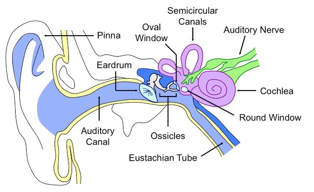
E.2.6 Label a diagram of the ear
E.2.7 Explain how sound is perceived by the ear, including the roles of the eardrum, bones of the middle ear, oval and round windows, and the hair . cells of the cochlea
- Sound travels as pressure waves in the air which push the membrane of the eardrum, causing it to vibrate
- The degree of vibration will vary according to the frequency and amplitude of the sound waves
- The ear drum pushes on the bones of the middle ear (the ossicles), which magnify the vibrations (~ 20 times)
- The bones of the middle ear are known as the hammer (malleus), anvil (incus) and stirrup (stapes)
- The ossicles push against the oval window, displacing fluid within the cochlea
- Movement of the cochlear fluid affects the position of cilia on sensory hair cells
- Cilia on hair cells vary in length and each resonates to a different frequency of sound
- Activation of the hair cells generates nerve impulses which are transmitted via the auditory nerve to the brain
- The kinetic motion of the cochlear fluid is dissipated by the movement of the round window