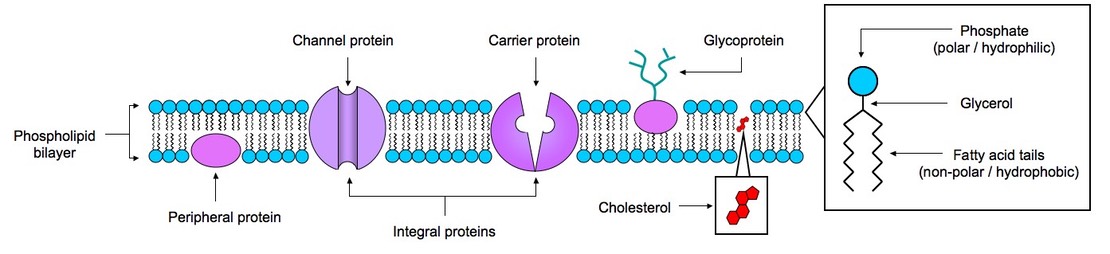
2.4.1 Draw and label a diagram to show the structure of membranes
2.4.2 Explain how the hydrophilic and hydrophobic properties of phospholipids help to maintain the structure of cell membranes
Structure of Phospholipids
- Consist of a polar head (hydrophilic) made from glycerol and phosphate
- Consist of two non-polar fatty acid tails (hydrophobic)
Arrangement in Membrane
- Phospholipids spontaneously arrange in a bilayer
- Hydrophobic tail regions face inwards and are shielded from the surrounding polar fluid while the two hydrophilic head regions associate with the cytosolic and extracellular environments respectively
Structural Properties of Phospholipid Bilayer
- Phospholipids are held together in a bilayer by hydrophobic interactions (weak associations)
- Hydrophilic / hydrophobic layers restrict entry and exit of substances
- Phospholipids allow for membrane fluidity / flexibility (important for functionality)
- Phospholipids with short or unsaturated fatty acids are more fluid
- Phospholipids can move horizontally or occasionally laterally to increase fluidity
- Fluidity allows for the breaking / remaking of membranes (exocytosis / endocytosis)
2.4.3 List the functions of membrane proteins

Transport: Protein channels (facilitated) and protein pumps (active)
Receptors: Peptide-based hormones (insulin, glucagon, etc.)
Anchorage: Cytoskeleton attachments and extracellular matrix
Cell recognition: MHC proteins and antigens
Intercellular joinings: Tight junctions and plasmodesmata
Enzymatic activity: Metabolic pathways (e.g. electron transport chain)
2.4.4 Define diffusion and osmosis
Diffusion:
The net movement of particles from a region of high concentration to a region of low concentration (along the gradient) until equilibrium
Osmosis:
The net movement of water molecules across a semi-permeable membrane from a region of low solute concentration to a region of high solute concentration until equilibrium is reached
![]() Osmosis
Osmosis
2.4.5 Explain passive transport across membranes in terms of simple diffusion and facilitated diffusion
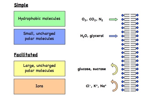
- The plasma membrane is semi-permeable and selective in what can cross
- Substances that move along the concentration gradient (high to low) undergo passive transport and do not require the expenditure of energy (ATP)
Simple diffusion:
- Small, non-polar (lipophilic) molecules can freely diffuse across the membrane
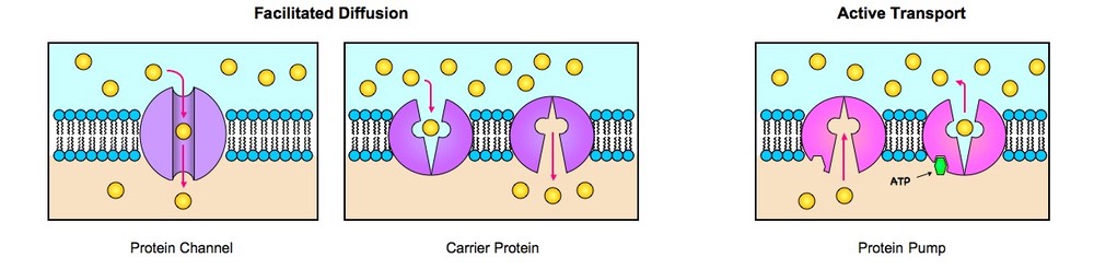
Facilitated diffusion:
- Larger, polar substances (ions, macromolecules) cannot freely diffuse and require the assistance of transport proteins (carrier proteins and channel proteins) to facilitate their movement (facilitated diffusion)
2.4.6 Explain the role of protein pumps and ATP in active transport across membranes
- Active transport is the passage of materials against a concentration gradient (from low to high)
- This process requires the use of protein pumps which use the energy from ATP to translocate the molecules against the gradient
- The hydrolysis of ATP causes a conformational change in the protein pump resulting in the forced movement of the substance
- Protein pumps are specific for a given molecule, allowing for movement to be regulated (e.g. to maintain chemical or electrical gradients)
- An example of an active transport mechanism is the Na+/K+ pump which is involved in the generation of nerve impulses
Types of Membrane Transport
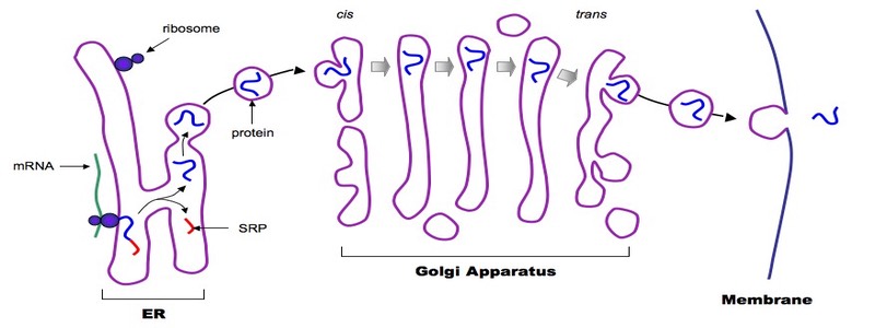
2.4.7 Explain how vesicles are used to transport materials within a cell between the endoplasmic reticulum, Golgi apparatus and plasma membrane
- Polypeptides destined for secretion contain an initial target sequence (a signal recognition peptide) which directs the ribosome to the endoplasmic reticulum
- The polypeptide continues to be synthesised by the ribosome into the lumen of the ER, where the signal sequence is removed from the nascent chain
- The polypeptide within the rough ER is transferred to the golgi apparatus via a vesicle, which forms from the budding of the membrane
- The polypeptide moves via vesicles from the cis face of the golgi to the trans face and may be modified along the way (e.g. glycosylated, truncated, etc.)
- The polypeptide is finally transferred via a vesicle to the plasma membrane, whereby it is either immediately released (constitutive secretion) or stored for a delayed release in response to some cellular signal (regulatory secretion = for a more concentrated and more sustained effect)
Overview of Vesicular Transport
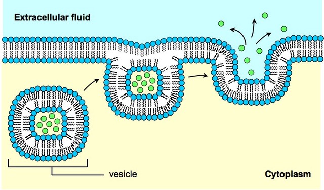
2.4.8 Describe how the fluidity of the membrane allows it to change shape, break and reform during endocytosis and exocytosis
The membrane is principally held together by the relatively weak hydrophobic associations between phospholipids
This association allows for membrane fluidity and flexibility, as the phospholipids (and to a lesser extent the proteins) can move about to some extent
This allows for the breaking and remaking of membranes, allowing larger substances access into and out of the cell (this is an active process)

Endocytosis
- The process by which large substances (or bulk amounts of smaller substances) enter the cell without travelling across the plasma membrane
- An invagination of the membrane forms a flask-like depression which envelopes the material; the invagination is then sealed off forming a vesicle
- There are two main types of endocytosis:
1. Phagocytosis
- The process by which solid substances (e.g. food particles, foreign pathogens) are ingested (usually to be transported to the lysosome for break down)
2. Pinocytosis
- The process by which liquids / solutions (e.g. dissolved substances) are ingested by the cell (allows quick entry for large amounts of substance)
Exocytosis
- The process by which large substances exit the cell without travelling across the plasma membrane
- Vesicles (usually derived from the golgi) fuse with the plasma membrane expelling their contents into the extracellular environment
The Process of Exocytosis