F.1.1 Outline the classification of living organisms into three domains
Living organisms were originally divided into five kingdoms based on the presence of certain structural features
In 1978 this system was refined to account for clear biochemical differences between living organisms (specifically, differences in rRNA sequence)
According to the three domain classification scheme, there are three distinct types of cellular organisms to which all living things may belong:
- Eukarya: Contain a membrane-bound nucleus (includes plants, animals, protists and fungi)
- Eubacteria: Lack a nucleus and consist of the traditional or 'true' bacteria (e.g. most pathogenic forms, E.coli, S. aureus, etc.)
- Archaea: Lack a nucleus and consist of the extremophiles or 'ancient' bacteria (e.g. methanogens, thermophiles, halophiles)
F.1.2 Explain the reasons for the reclassification of living organisms into three domains
- Traditional classification schemes separated organisms into two groups: prokaryotes and eukaryotes
- The large diversity of the group categorised as prokaryotes prompted further division into two separate domains
- Differences in the genes that transcribed rRNA as well as certain structural features (e.g. cell wall composition) formed the basis for this separation
- The archaebacteria have been found to have certain features that share more in common with eukaryotes that eubacteria
- The reclassification of organisms into three domains has helped scientists to better study and understand the origin and evolution of eukaryotes
F.1.3 Distinguish between the characteristics of the three domains
Each of these three domains (Eukarya, Eubacteria, Archaea) contain rRNA which is unique to them and forms the basis of their division

F.1.4 Outline the wide diversity of habitat in the Archaea as exemplified by methanogens, thermophiles and halophiles
Methanogens
- Obligate anaerobes (cannot survive in the presence of oxygen)
- Produce methane as a waste product and are found in marshes, the guts of animals and oxygen depleted soils (e.g. landfills)
Thermophiles
- Can survive abnormally high temperatures and live at temperatures close to boiling (60 – 100ºC)
- Are found in hot springs and near geothermal lava flows (e.g. deep sea vents and volcanic calderas)
Halophiles
- Live in saline habitats with very high salt concentrations
- Found in regions of the Great Salt Lake and the Dead Sea
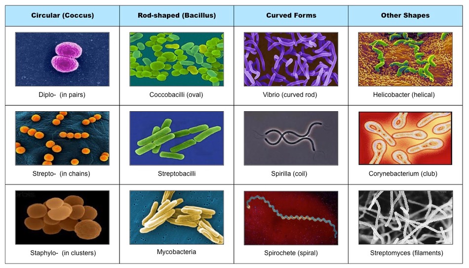
F.1.5 Outline the diversity of Eubacteria, including shape and cell wall structure
Eubacteria can be sub-classified according to a number of diverse features, including:
- Shape: Round (coccus), rod-shaped (bacillus), comma-shaped (vibrio) or spiral (spirilla / spirochete)
- Cell wall composition: Gram positive (thick peptidoglycan layer) or Gram negative (lipopolysaccharide layer)
- Gaseous requirements: Anaerobic (obligate vs facultative) or aerobic
- Nutritional patterns: Autotrophic (photosynthetic vs chemosynthetic) or heterotrophic
- Additional structures: Flagella, slime capsules, hyphae, endospores, etc.
Types of Eubacteria According to Shape
F.1.6 State, with one example, that some bacteria form aggregates that show characteristics not seen in individual bacteria
- Bacteria may form aggregates and by interacting are capable of completing functions that individual cells could not undertake (emergent properties)
- The photobacterium Vibrio fischeri is able to emit light (biolouminescence) when they become part of a population with high density
- V. fischeri releases a regulatory substance into its surroundings which in dense populations becomes concentrated enough to trigger bioluminescence
F.1.7 Compare the structure of the cell walls of Gram-positive and Gram-negative Eubacteria
Similarities:
- Both have walls made of a murein net
- Both contain peptidoglycan
Differences:
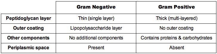
Gram Negative versus Gram Positive Cell Walls

F.1.8 Outline the diversity of structure in viruses including: naked capsid vs enveloped capsid; DNA vs RNA and single vs double-stranded DNA or RNA
- A virus is a non-cellular agent consisting of a protein coat (capsid) and genetic material
- The genetic material may be DNA (adenovirus) or RNA (retrovirus) and may be single-stranded or double-stranded
- For some viruses the protein coat may be exposed (naked capsid) while others may be covered in a membranous bilayer (enveloped capsid)
- Retroviruses have a reverse transcriptase component to allow for production of viral DNA
Types of Viruses (Not to Scale)

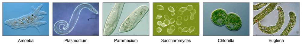
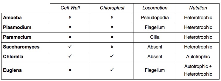
F.1.9 Outline the diversity of microscopic eukaryotes, as illustrated by Saccharomyces, Amoeba, Plasmodium, Paramecium, Euglena and Chlorella
- Amoeba – single-celled organism that inhabits freshwater ponds and moves and captures food via cell extensions called pseudopodia
- Plasmodium – a genus of parasitic protozoa that causes malaria as part of a complex life cycle involving two hosts (humans and mosquitoes)
- Paramecium – a protozoan that inhabits freshwater environments and moves via the coordinated beating of tiny cilia
- Saccharomyces – a genus of single-celled fungus known as yeasts and employed extensively in the fermentation process
- Chlorella – a genus of unicellular green algae that possesses a singular cup-shaped chloroplast within its cytoplasm
- Euglena – a flagellated protozoan that contains chloroplast and is commonly present in pond water
Key Structural and Functional Features of Microscopic Eukaryotes
Overview of Microscopic Eukaryotes