E.5.1 Label, on a diagram of the brain, the medulla oblongata, cerebellum, hypothalamus, pituitary gland and cerebral hemispheres
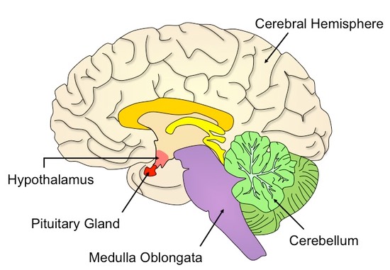
E.5.2 Outline the functions of each of the parts of the brain listed in E.5.1
Medulla oblongata: Controls automatic and homeostatic activities, such as swallowing, digestion and vomiting, and breathing and heart rate
Cerebellum: Coordinates unconscious functions, such as movement and balance
Hypothalamus: Maintains homeostasis via coordination of the nervous and endocrine systems, produces hormones secreted by posterior pituitary
Pituitary Gland: Produces and secretes hormones regulating many body functions - such as ADH (water retention / osmoregulation)
Cerebral Hemispheres: Acts as the integration centre for highly complex functions, such as learning, memory and emotion
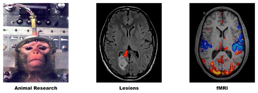
E.5.3 Explain how animal experiments, lesions and fMRI scanning can be used in the identification of the brain part involved in specific functions
Animal Experiments
- One method of investigating the function of specific brain regions is to either stimulate the region with electrodes or remove the region (lobotomy) and assess the physical effect
- Because such procedures are highly invasive and potentially damaging, animal models are frequently used
- Such studies may be limited by the fact that animal brains may be dissimilar to human brains, so valid comparisons can be hard to make
- The best comparisons are made with primates, however ethical considerations are of greater concern with these models
- Animal experimentation using rats and mice have been used to understand and develop drug treatments for brain diseases such as MS
Lesions
- Lesions are abnormal areas in brain tissue (either from accidents or congenital) which can indicate the effect of the loss of an area
- Such studies may be limited by the fact that many functions involve multiple brain areas, making complex effects difficult to interpret
- The brain also has the ability to re-learn skills by re-routing the function through another part of the brain (plasticity)
- Split brain patients (severed corpus callosum) have been used to identify the functional roles of the left and right hemispheres
- Individuals with aphasia (impairment to use or understand language) often have damage to specific regions of the left hemisphere
Functional Magnetic Resonance Imaging (fMRI)
- fMRI records changes in blood flow and can indicate active regions of the brain via an increase in blood flow
- Oxygenated haemoglobin (oxyhaemoglobin) responds differently to a magnetic field than deoxygenated haemoglobin
- These differences in oxygenation (reflecting level of brain activity) can be represented visually by different colours
- When a subject is given a stimulus designed to stimulate specific brain activity, fMRI can be used to localise regions involved
- Temporal activities can also be recorded to allow identification of sequential collaboration between brain parts
- While this procedure is non-invasive and can be performed on healthy subjects, not all brain activity is detected by fMRI
- fMRI studies have been used to diagnose ADHD and dyslexia, in addition to being used to monitor recovery from strokes
Methods of Identification of Brain Regions Involved in Specific Functions
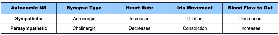
E.5.4 Explain sympathetic and parasympathetic control of the heart rate, movement of the iris and flow of blood to the gut
The sympathetic and parasympathetic nervous systems are part of the autonomic system and have antagonistic actions
Sympathetic Nervous System
- Involved in processes that prepare the body for action ('fight or flight' responses)
- The sympathetic nervous system releases noradrenaline (adrenergic) via sympathetic nerves
- Heart rate will increase to improve blood flow (via release of adrenaline)
- Pupils will dilate to improve vision (via contraction of radial muscles)
- Blood flow to gut will decrease (due to vasoconstriction of blood vessels by smooth muscles)
Parasympathetic Nervous System
- Involved in processes that occur when the body is relaxed ('rest and digest' or 'feed and breed' responses)
- The parasympathetic nervous system releases acetylcholine (cholinergic) via parasympathetic nerves
- Heart rate will decrease to reduce blood flow (via stimulation by vagus nerve)
- Pupils will constrict to restrict light and potential retinal damage (via contraction of circular muscles)
- Blood flow to gut will increase to facilitate digestion (due to vasodilation of blood vessels by smooth muscles)
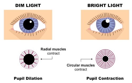
E.5.5 Explain the pupil reflex
- The pupil reflex is a cranial reflex originating at the brainstem and under the control of the autonomic nervous system
- Bright light can overstimulate photoreceptors and potentially damage the retina
- In changing levels of light, the iris will automatically resize to regulate the amount of light that reaches the retina
In dim light conditions:
- Relay neurons stimulate the sympathetic nervous system, causing radial muscles to contract and the pupil to dilate
In bright light conditions:
- Relay neurons stimulate the parasympathetic nervous system, causing circular muscles to contract and the pupil to constrict
E.5.6 Discuss the concept of brain death and the use of the pupil reflex in testing for this
- Whole brain death is the permanent absence of measurable activity in both the cerebrum and the brain stem
- The cerebrum is involved in higher order brain functions, while the brain stem may function alone to maintain homeostasis
- Individuals with a non-functioning cerebrum but functioning brain stem may be kept alive in a vegetative state
- Without a functioning brain stem, life cannot continue as homeostatic processes cannot continue to function
- Failure of a pupil to respond to light indicates brain stem death and thus the pupil reflex is used to test for whole brain death
E.5.7 Outline how pain is perceived and how endorphins can act as painkillers
- Pain is perceived when impulses pass from pain receptors (nocireceptors) in body tissues to sensory areas of the cerebral cortex
- Endorphins are released by the pituitary gland during stress, injury or exercise and act as painkillers by blocking pain perception
- They do this by blocking the release of neurotransmitters at the synapses involved in pain signal transmission