H.2.1 State that digestive juices are secreted into the alimentary canal by glands, including salivary glands, gastric glands in the stomach wall, the pancreas and the wall of the small intestine
Chemical digestion in the alimentary canal involves the secretion of digestive juices capable of breaking down complex macromolecules
These digestive juices are secreted from glands, which include:
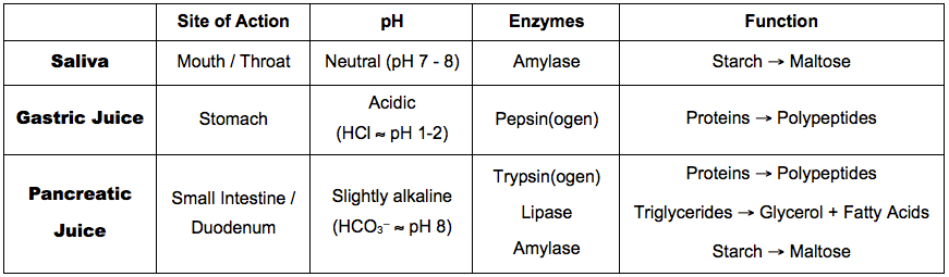
- Salivary glands – secretes saliva which contains amylase
- Gastric glands – secretes gastric juices which include hydrochloric acid and pepsinogen
- Pancreas – secretes pancreatic juices containing lipase, trypsinogen and hydrogen
- Intestinal wall – The small intestine contains the crypts of Lieberkuhn, which secrete a variety of substances as part of the intestinal juice
H.2.2 Explain the structural features of exocrine gland cells
- Exocrine glands have ducts through which they secrete their product (these ducts may arise from the convergence of smaller ductules)
- The ducts / ductules arise from a cluster of cells called an acinus (plural = acini), surrounded by a basement membrane
- Acini are lined by a single layer of secretory cells which release the exocrine product into the lumen of the duct via secretory vesicles
- Secretory cells are held together by tight junctions, and possess a highly developed rough ER and golgi network for material secretion
Structure of an Exocrine Gland (Unbranched Tubular)

H.2.3 Compare the composition of saliva, gastric juice and pancreatic juice
Similarities:
- Contain water (universal solvent)
- Contain mucus (as a lubricant or an intestinal lining)
- Contain salts and ions (calcium, phosphate, etc.)
Differences:
H.2.4 Outline the control of digestive juice secretion by nerves and hormones, using the example of secretion of gastric juice
Pre-Ingestion
- The sight and smell of food triggers a reflex response in which gastric juice is secreted from gastric pits in the stomach wall
- This ensures that gastric juice is in the stomach by the time the food is consumed
Post-Ingestion
- Food entering the stomach causes distension, which is detected by stretch receptors in the stomach lining
- Impulses are sent to the brain, which triggers the secretion of gastrin from the pits lining the stomach wall
- Gastrin causes the sustained release of gastric juice, particularly its acid component
- When the pH drops too low, gastrin secretion is inhibited by hormones (secretin and somatostatin)
H.2.5 Outline the role of membrane-bound enzymes on the surface of epithelial cells in the small intestine in digestion
Some digestive enzymes are immobilised on the plasma membrane of the epithelial cells of the small intestine, serving two main benefits:
1. The enzyme is fixed in place and does not pass through the digestive system, meaning it can be reused
2. The enzyme can be linked to secondary functions (e.g. membrane transport)
Example:
- Maltase is immobilised on the epithelial lining with its active site facing towards the intestinal lumen
- Maltase digests the disaccharide maltose into two glucose monomers, which are then absorbed by localised transporters
H.2.6 Outline the reason for cellulose not being digested in the alimentary canal
Glucose can exist in one of two isomeric forms: a-glucose or ß-glucose
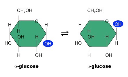
While humans can digest polymers of a-glucose (e.g. starch, glycogen), they cannot digest the polymer of ß-glucose (cellulose)
- This is because they do not produce the necessary enzyme (cellulase) and lack bacteria in their gut capable of digesting cellulose
- Cellulose is a component of plant cell walls and the main source of dietary roughage (undigested, it creates bulk which stimulates peristalsis)
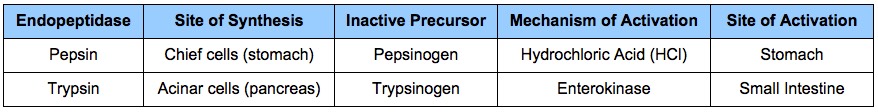
H.2.7 Explain why pepsin and trypsin are initially synthesised as inactive precursors and how they are subsequently activated
- Both pepsin and trypsin are protease enzymes (specifically endopeptidases) which hydrolyse peptide bonds to digest proteins
- As proteins are ubiquitous and essential components of cells, these enzymes could digest the cells that secrete them (auto digestion)
- Instead, they are synthesised as inactive forms (zymogens) and subsequently activated in the digestive tract (lined with mucus to protect cells)
Activation of Endopeptidases
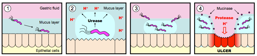
H.2.8 Discuss the roles of gastric acid and Helicobacter pylori in the development of stomach ulcers and stomach cancers
- Stomach ulcers are inflammed and damaged areas in the stomach wall
- There is a strong correlation between H. pylori infection and the development of stomach ulcers
- H. pylori is a bacterium that can survive the acid conditions of the stomach
- It secretes urease which neutralises the gastric acid to lower the acidity of the stomach for further colonisation
- It also secretes proteases (e.g. mucinase) to degrade the mucosal lining of the stomach wall, allowing it to burrow into this lining
- The degradation of this protective lining by H. pylori allows for damage to the stomach wall by gastric acids (causing ulcers)
- The prolonged presence of stomach ulcers may lead to the formation of stomach cancers
- There is a correlation between chronic H. pylori infection over a number of years (20 - 30 years) and the development of stomach cancer
- The link between H. pylori and development of stomach ulcers / cancers represents a paradigm shift – cause was previously thought to be stress
Stages of Helicobacter pylori Infection
H.2.9 Explain the problem of lipid digestion in a hydrophilic medium and the role of bile in overcoming this
- Lipids are hydrophobic and hence insoluble within the aqueous environment of the body
- They will group together (coalesce) to form large droplets of fat (fat globules)
- The enzyme responsible for lipid digestion (lipase) is water soluble and can only bind the lipids to its active site at the lipid-water interface (i.e. the surface of the fat globule)
- As the interior of the fat globule is inaccessible to lipase in this form, this would make lipid digestion normally very slow

- Bile is a watery fluid that contains bile salts and pigments (bilirubin) – it is produced by liver cells and stored in the gall bladder
- Bile salt molecules have both a hydrophobic and hydrophilic end
- The hydrophobic end attaches to the lipid while the hydrophilic end interacts with water – preventing lipids from attaching there
- This divides the fat globule into smaller droplets (emulsification), increasing the total surface area available for enzyme activity
![]() Lipid Absorption
Lipid Absorption