
Non-Disjunction
Sometimes errors may occur during the process of cell division, resulting in defective cells that can contribute to certain disease conditions
-
Some errors maybe prevent cells from completing cell division, resulting in the gradual degeneration of the tissue (e.g. Alzheimer’s disease)
-
Other errors may cause cell division to become uncontrolled, leading to the unregulated proliferation of cells (e.g. cancers)
Non-Disjunction
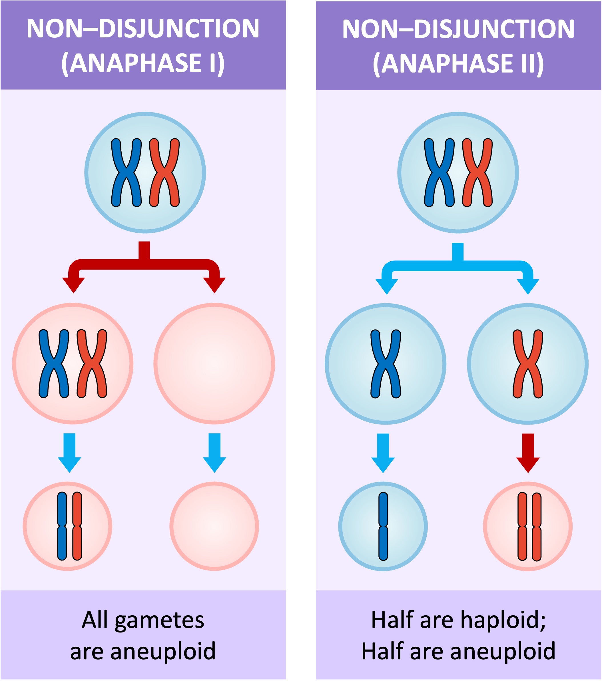
Non-disjunction is a nuclear division error whereby specific chromosomes fail to separate correctly during anaphase
-
When non-disjunction occurs during meiosis, the resulting gamete will possess either an extra, or a missing, copy of a chromosome
-
If a zygote is formed from that gamete, then the resulting offspring will have an incorrect chromosome number in every cell of their body
Non-disjunction can occur during either Anaphase I (affecting all four daughter cells) or Anaphase II (only two of the four daughter cells will be affected)
-
The presence of an abnormal chromosome number in a cell is known as aneuploidy (‘aneu’ = without, ‘ploidy’ = chromosome number)
Meiotic Non-Disjunction


Studies show that the chances of non-disjunction increase as the age of the parents increase
-
There is a particularly strong correlation between maternal age and the occurrence of non-disjunction, with the risk of chromosomal abnormalities in offspring increasing significantly after a maternal age of 30
-
The other parental gamete was normal and had a single copy of chromosome 21
-
This may be due to developing oocytes being arrested in prophase I until ovulation as part of the process of oogenesis
Down Syndrome
A common condition that arises from a non-disjunction event is Down syndrome (three copies of chromosome 21)
-
One of the parental gametes had two copies of chromosome 21 as a result of non-disjunction
-
The other parental gamete was normal and had a single copy of chromosome 21
-
When the two gametes fused during fertilisation, the resulting zygote had three copies (trisomy) of chromosome 21
Down syndrome can be detected via a process known as karyotyping, by which chromosomes are organised and visualised for inspection
-
Cells are harvested from the foetus before being chemically induced to undertake cell division (so chromosomes are visible)
-
The chromosomes are stained and photographed, before being organised (according to size) to generate a visual profile called a karyogram
-
Foetal cells can be harvested from the placenta (chorionic villi sampling) or the amniotic fluid (amniocentesis)
-
Placental cells can be harvested sooner but carry a slightly higher risk of miscarriage
-
Female with Down Syndrome


