
Microscopes
Microscopes are scientific instruments that are used to visualise objects that are too small to see with the naked eye
-
There are two main types of microscope – optical (light) microscopes and electron microscopes
Light Microscopy
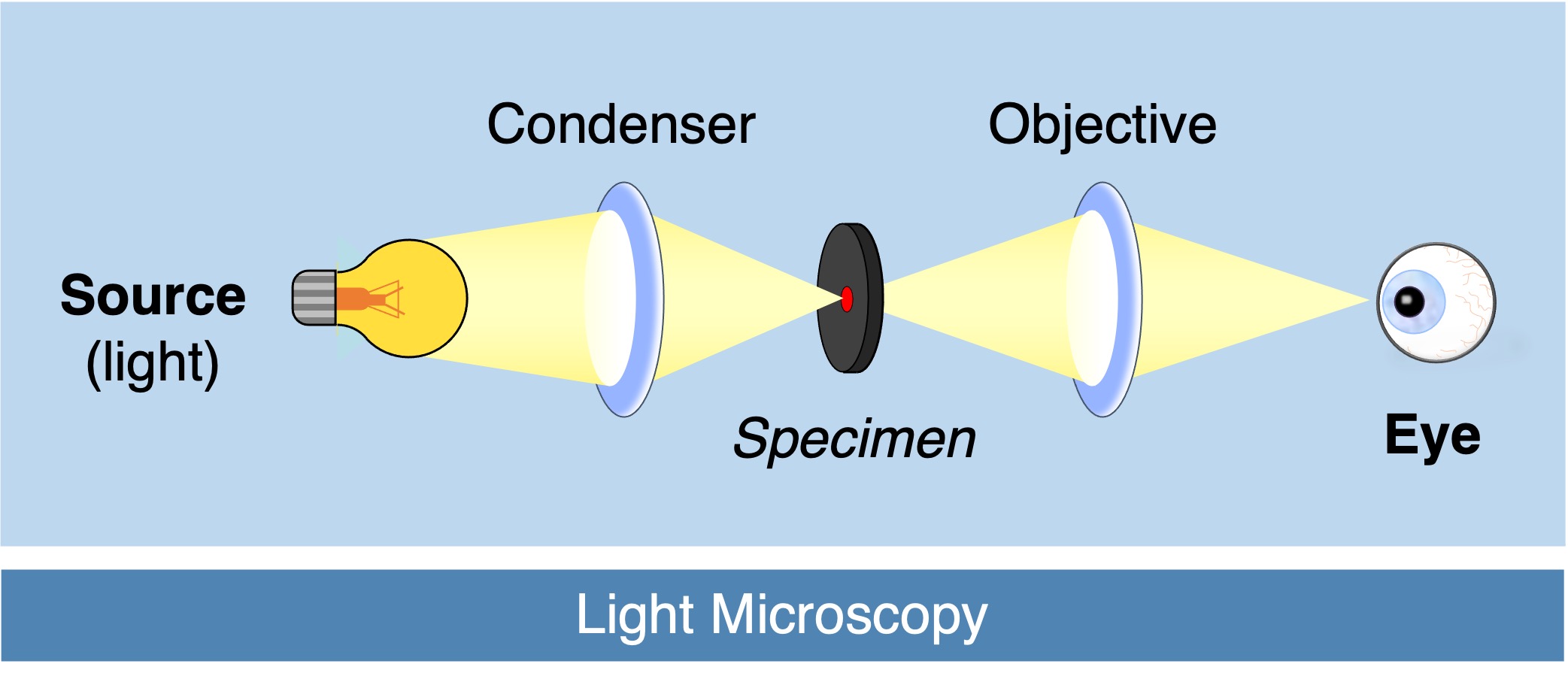
Light microscopes can be used to view living specimens in their natural colours
-
These microscopes use glass lenses to bend light in order to magnify images (extent of magnification is determined by the lenses used)
The clarity of cellular sub-structures can be improved via the use of fluorescent labelling
-
Synthetic dyes can be used to bind particular cellular compounds in order to resolve specific structures (e.g. DAPI is a fluorescent dye that stains DNA)
-
Immunofluorescence staining uses antibodies that are conjugated to fluorescent probes to specifically target a cellular component of choice
Light Microscope Overview


Electron Microscopy
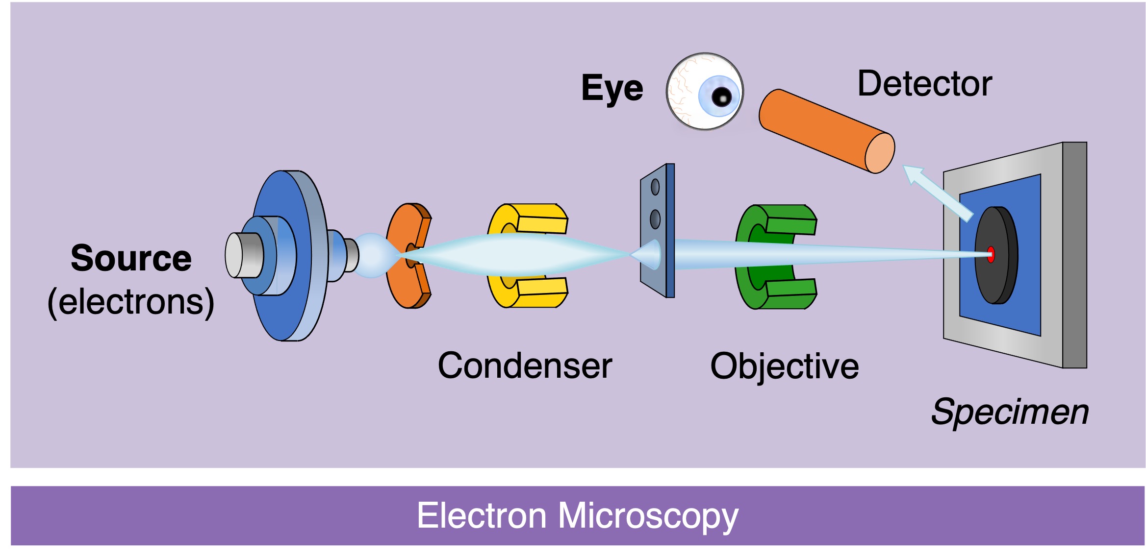
Electron microscopes can generate images at a much higher magnification and resolution, however cannot view living specimens in natural colour
-
These microscopes use electromagnets to focus electrons and produce monochromatic images (to which false colour may be applied)
There are two main types of electron microscopes that allow for different visual representations of a biological specimen
-
Transmission electron microscopes (TEMs) pass electrons through a specimen to generate a cross-section image
-
Scanning electron microscopes (SEMs) scatter electrons over a surface to differentiate depth and map in 3D
Cryogenic electron microscopy involves freezing samples prior to viewing in order to generate images of a comparable standard to X-ray crystallography
-
This allows for the determination of molecular structures at near atomic resolution without requiring the crystallisation of the specimen
-
If the frozen specimen is cracked along a plane via freeze fracturing, then internal cellular structures can be studied
-
Freeze fracturing was used to demonstrate the presence of integral membrane proteins within the plasma membrane
Electron Microscope Overview


