
Microscopy Skills
Light microscopes use visible light and a combination of lenses to magnify images of mounted specimens
-
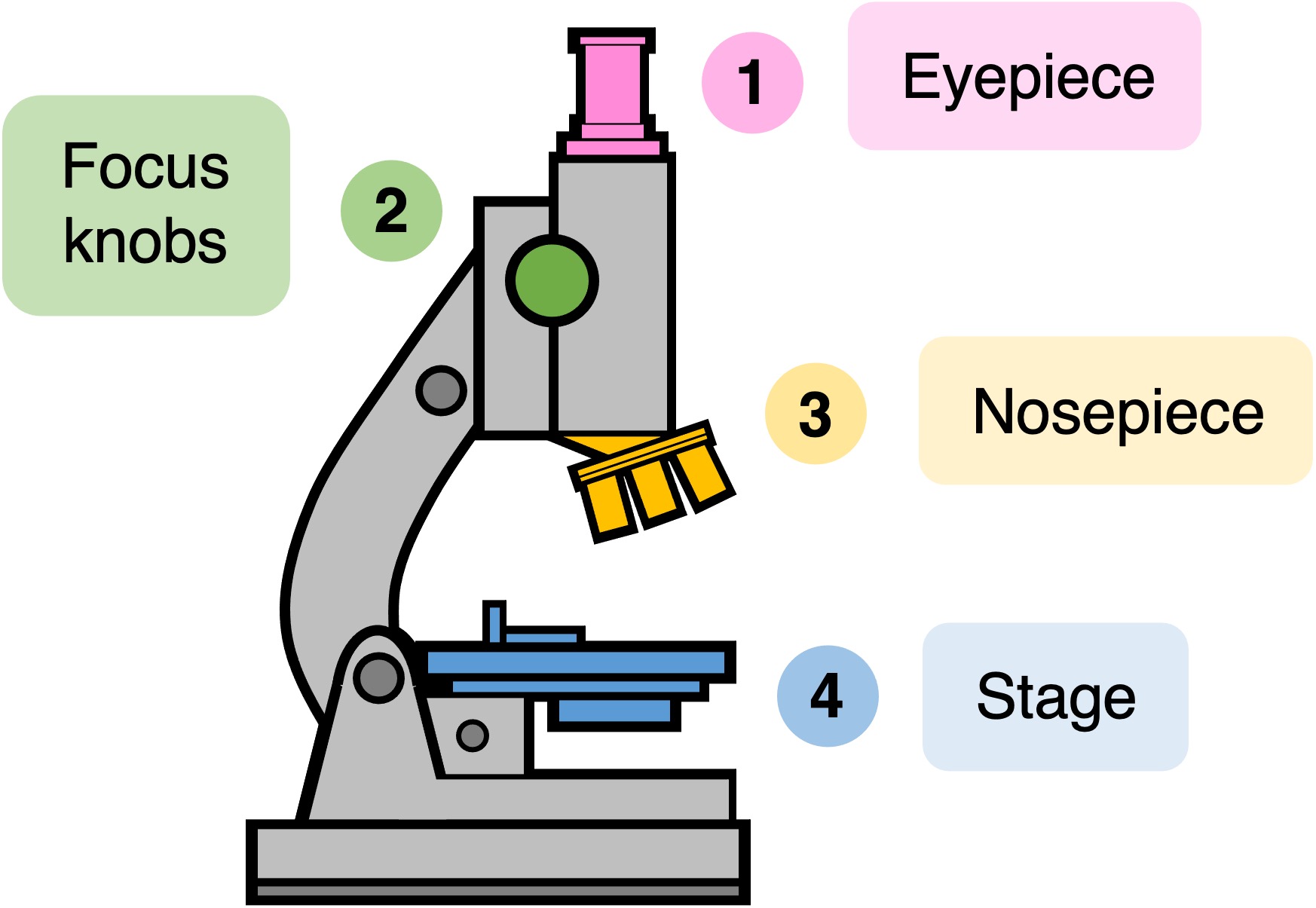
Most light microscopes include both an ocular lens (~10×) and objective lens (~10×; 40×; 100×)
Using a Light Microscope
When using a light microscope to view biological specimens, the following conventions should be followed:
-
The image should initially be resolved at the lowest magnification using the coarse focus mechanism
-
Higher magnifications are then obtained by changing the objective lens (via the revolving nosepiece) and making fine focus adjustments
-
The total magnification of the image is calculated by multiplying the magnification of both lenses (ocular and objective) together
-
If the eyepiece has a measurement scale on its surface (eyepiece graticule), this can be used to determine sizes of biological structures
Light Microscope Components


Calculating Magnification
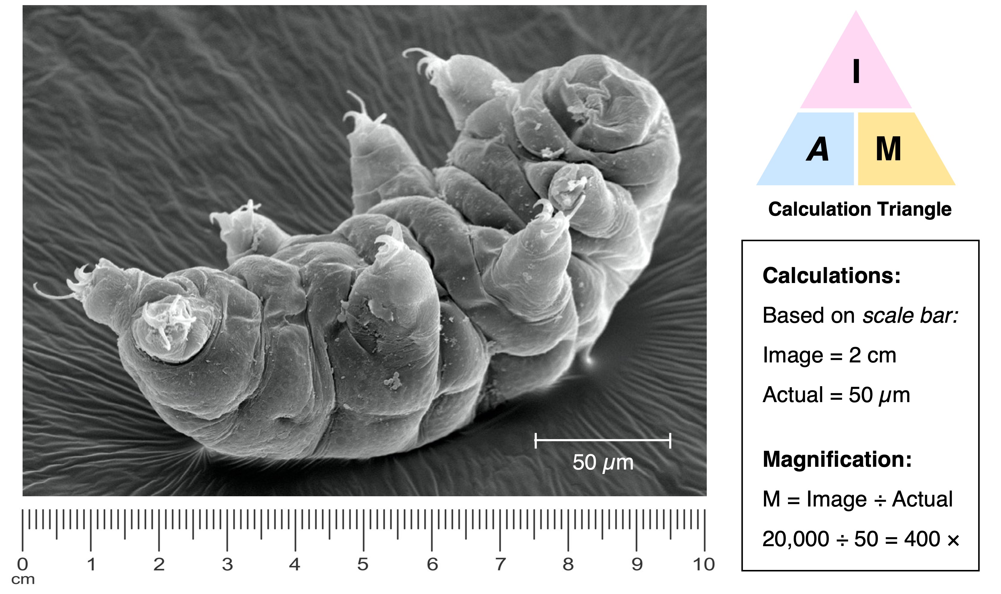
To calculate the linear magnification of a drawing or image, the following equation should be used:
-
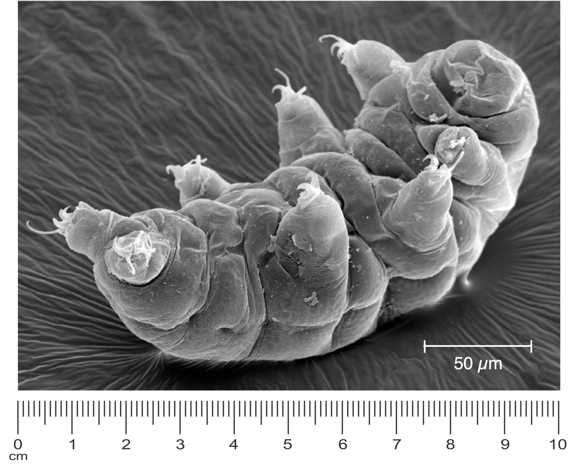
Magnification = Image size (with ruler) ÷ Actual size (according to scale bar)
In order to calculate magnification, both image size and actual size must be in the same units
-
Metric conversions can be applied according to the following table:
-
Unit
Metre
Centi-
Milli-
Micro-
Nano-
-
Sign
m
cm
mm
μm
nm
-
Size
1 (100)
10–2
10–3
10–6
10–9
-
Unit
Sign
Size
-
Metre
m
1 (100)
-
Centi-
cm
10–2
-
Milli-
mm
10–3
-
Micro-
μm
10–6
-
Nano-
nm
10–9
Example Calculation


