
Cell Adaptations
Cells that are specialised for material exchange will possess adaptations to increase their surface area
-
Cells that are flat and long (squamous) will have a higher SA:Vol ratio than cells that are larger or cuboidal in shape
-
Tissues lining absorption surfaces will have ruffled projections (villi), while the cells themselves will possess membranous extensions (microvilli)
Examples of cells that are specialised for material transport include:
-
Red blood cells: The cells are thin and flat (biconcave) and have had their nuclei removed to store more haemoglobin (increases oxygen uptake)
-
Tubule cells: The proximal convoluted tubule of the kidney is folded into villi and the tubule cells possess microvilli to improve selective reabsorption
SA:Vol Ratio Adaptations
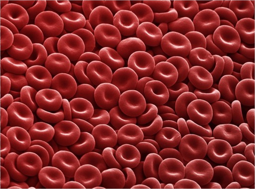
Red Blood Cells
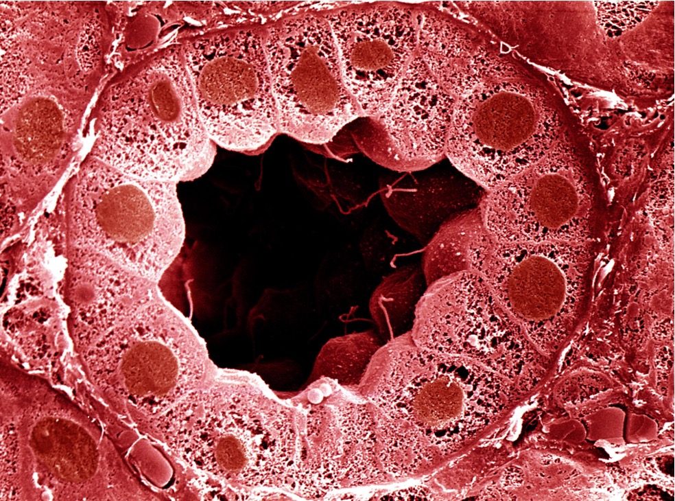
Kidney Tubule (Villi)
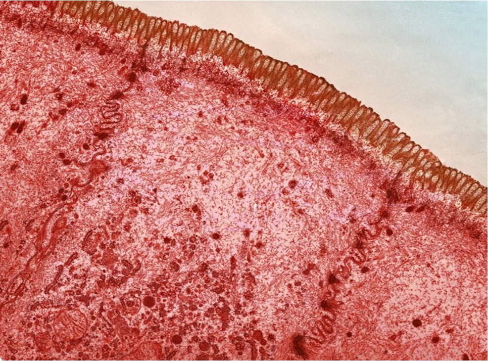
Tubule (Microvilli)
Pneumocytes
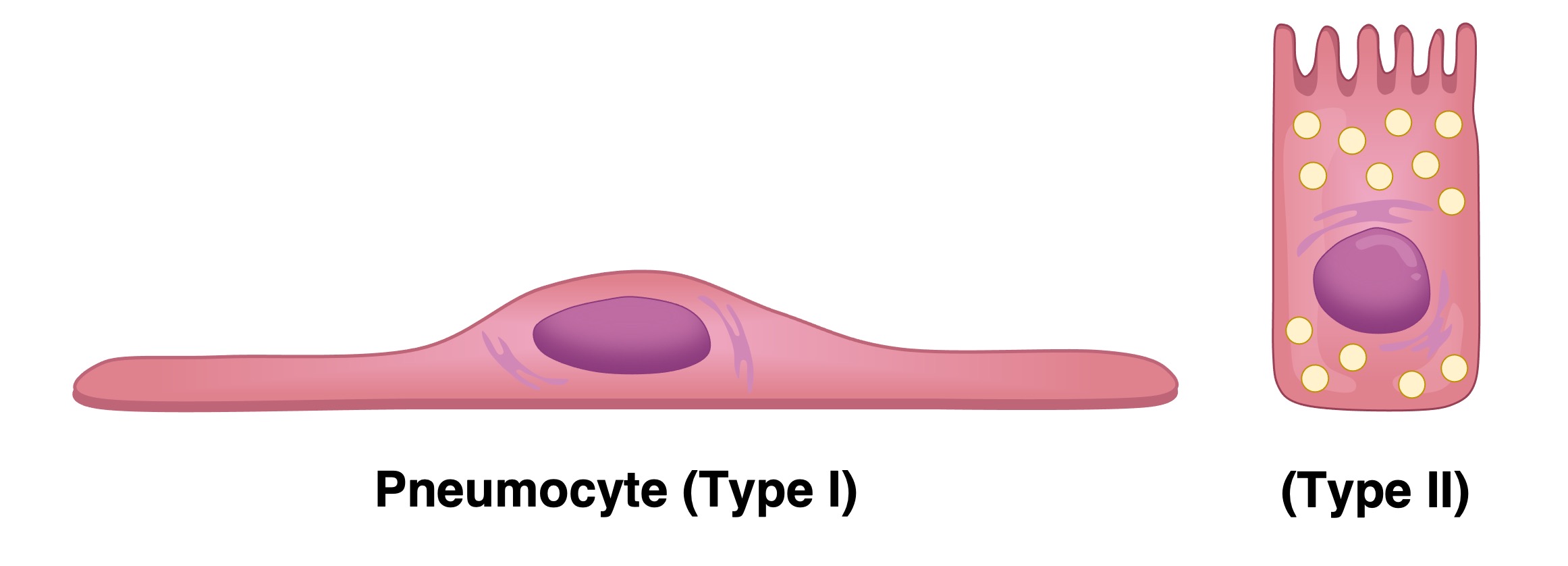
Pneumocytes (or alveolar cells) are the cells that line the alveoli and comprise of the majority of the inner surface of the lungs
-
There are two types of alveolar cells – type I pneumocytes and type II pneumocytes
Type I Pneumocytes:
-
Type I pneumocytes are involved in the process of gas exchange between the alveoli and the capillaries
-
They are squamous (flattened) in shape and extremely thin (~ 0.15µm) – minimising the diffusion distance for respiratory gases
-
The cells are connected by occluding junctions, which prevents the leakage of tissue fluid into the alveolar air space
Type II Pneumocytes:
-
Type II pneumocytes secrete pulmonary surfactant, which reduces surface tension in the alveoli (making it easier for them to inflate)
-
They are cuboidal in shape and possess many granules called lamellar bodies (which store the surfactant)
Types of Alveolar Cells
Muscles
All muscles contain long protein filaments (myofibrils) which are responsible for muscle contraction and relaxation – however different types of muscles possess specialised features according to their function
-
Striated muscles connect to the bones of the skeleton and are responsible for locomotion (voluntary movement)
-
Cardiac muscle cells are found within the heart tissue and are responsible for the rhythmic beating of the heart
Striated Muscle Fibres:
-
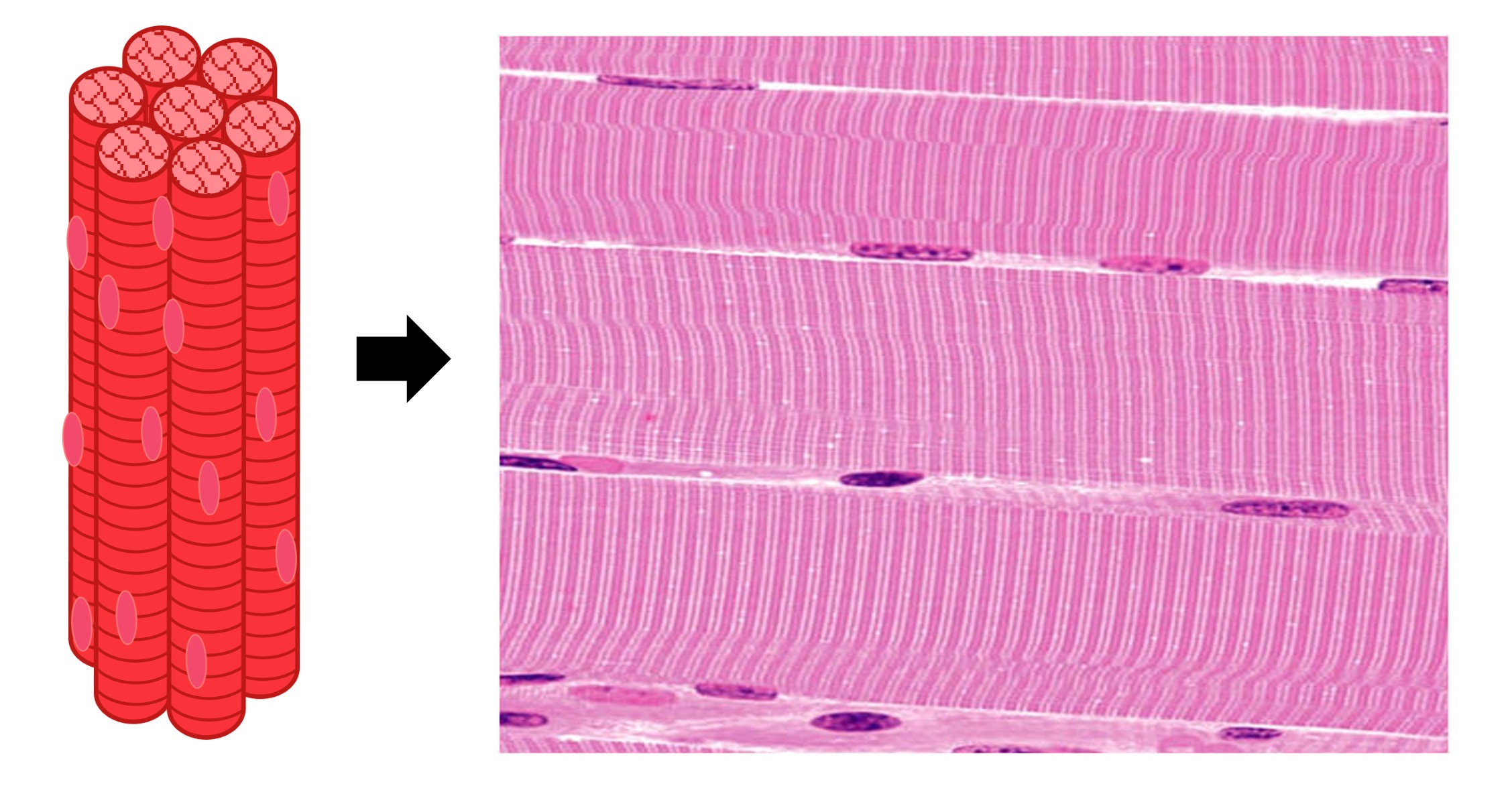
Skeletal muscles are comprised of long, cylindrical fibres that are formed from the fusion of individual cells
-
The fibres therefore have a single, continuous plasma membrane (sarcolemma) and are multinucleate
-
These long and cylindrical fibres are packed together in unbranching strands (typically 2–3 cm in length) that collectively form a muscle bundle
-
While the muscle fibre operates as a single functional unit, the fact that it was formed from the fusion of individual muscle cells makes it debatable whether a muscle fibre can be considered a cell
Cardiac Muscle Cells:
-
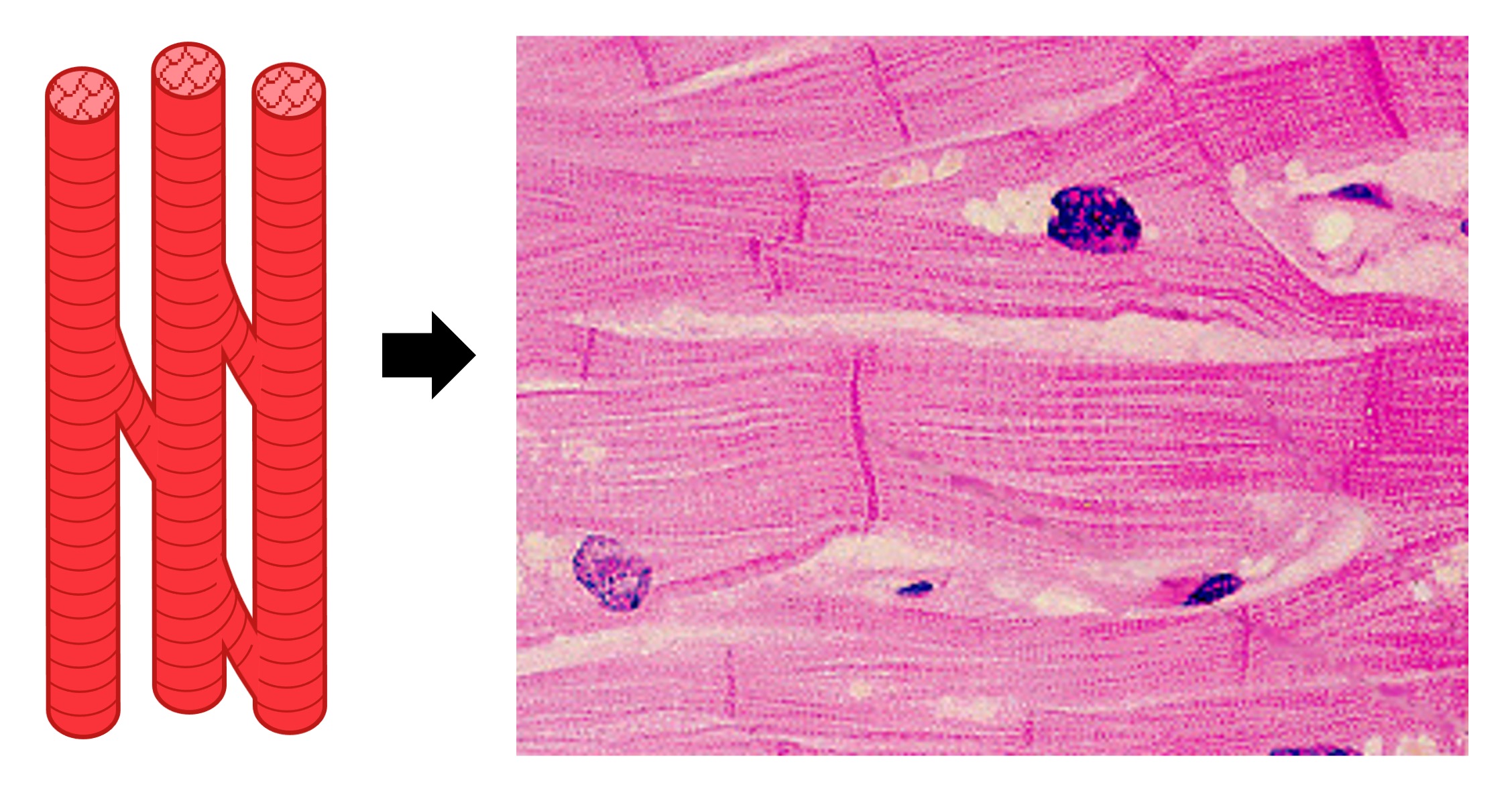
Cardiac muscle cells are short (~0.1 mm), narrow and fairly rectangular in shape
-
Unlike in skeletal muscle, the cardac muscle cells are not fused together and consequently are mononucleated (one nucleus per cell)
-
The individual muscle cells are connected by gap junctions at intercalated discs, which allow for electrical conduction between the cells
-
Cardiac muscle cells are branched, allowing for faster signal propagation and contraction in three dimensions
-
The cells have more mitochondria than striated muscle fibres as they are more reliant on aerobic respiration for ATP production
Types of Muscle
Skeletal Muscle
Cardiac Muscle
Gametes
The male and female reproductive gametes (sperm and egg) have specialised structures which reflect their functions
-
The male gamete (sperm) is small and motile and only contributes the male’s haploid nucleus to the zygote
-
The female gamete (egg) is large and non-motile and contributes all the organelles and cytoplasm to the zygote
Sperm:
-
A typical human spermatozoa can be divided into three sections – head, mid-piece and tail
-
The head region contains three structures – a haploid nucleus, an acrosome cap and paired centrioles
-
The haploid nucleus contains the paternal DNA (this will combine with maternal DNA upon fertilisation)
-
The acrosome cap contains hydrolytic enzymes which help the sperm to penetrate the egg
-
The centrioles are needed by a zygote to divide (egg cells expel their centrioles within their polar bodies)
-
-
The mid-piece contains high numbers of mitochondria which provide the energy (ATP) needed for the tail to move
-
The tail (flagellum) is composed of a microtubule structure called the axoneme, which bends to facilitate movement
Ovum (Egg):
-
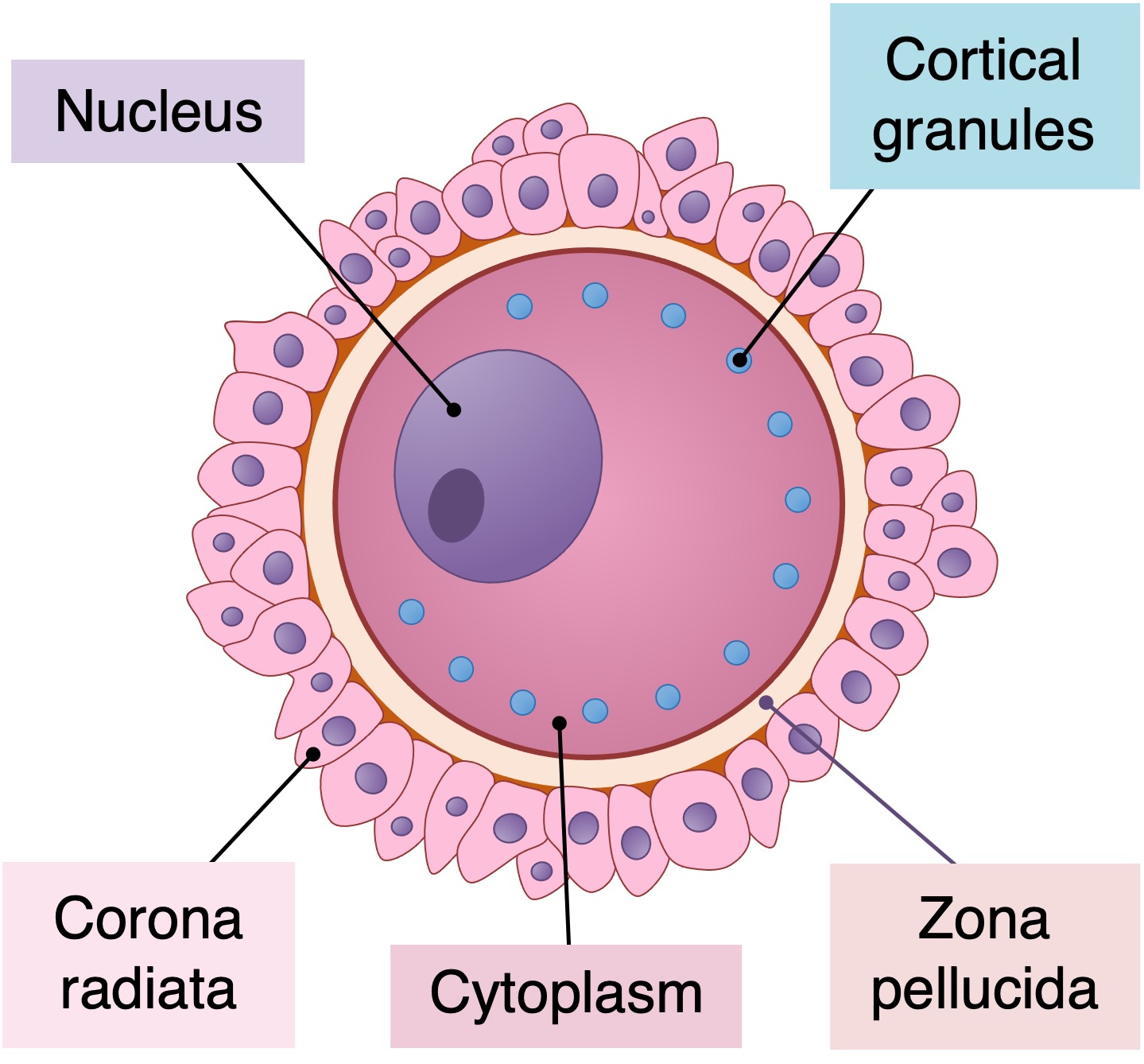
A typical egg cell is surrounded by two distinct layers – the zone pellucida (jelly coat) and corona radiata
-
The zona pellucida is a glycoprotein matrix which acts as a barrier to sperm entry
-
The corona radiata is an external layer of follicular cells which provide the egg with nourishment
-
-
Within the egg cell are numerous cortical granules, which release their contents upon fertilisation to prevent polyspermy
-
Although diagrams of egg cells commonly include a haploid nucleus, no nucleus will form within the egg until after fertilisation has occurred
-
The egg cell is arrested in metaphase II until it becomes fertilised by a sperm
-
Human Sperm and Egg


Human Sperm
